 LINE
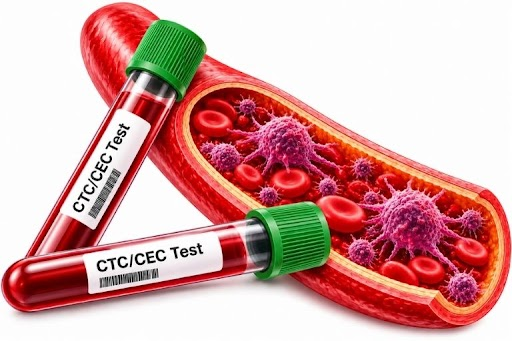
THE SHORT ANSWER
CEC are circulating endothelial cells; tCEC are studied in a tumour-associated context. Neither term is the same as CTC, and finding cells alone does not establish a cancer diagnosis. [1] [3]
Article information
These fields are unfilled for the responsible team. They do not indicate completed medical review or approval.
Written by
Scientific / medical reviewer
Approved by
What are CEC and tCEC?
Circulating endothelial cells (CEC) are endothelial cells found in the bloodstream. They may arise from established blood vessels or other endothelial-related sources. [1]
Tumour-associated circulating endothelial cells (tCEC) are studied in a tumour context; interpretation depends on the method and criteria. Circulating tumour cells (CTC) are tumour-derived cells in blood, a distinct group. [3]
Why is finding rare cells challenging?
Cells of interest may be scarce relative to other blood cells. Sample preparation, cell preservation and appropriate identification criteria matter because different cells can share characteristics. [2]
How does X-ZELL Cryoimmunostaining™ support analysis?
Cryoimmunostaining™ combines cold fixation and antibody staining to display information about selected targets in cell images. [4] [5]
X-ZELL describes Cryosuite as a workflow using fixation and staining instruments, reagents and multichannel imaging. The actual protocol and panel depend on the sample and laboratory method. [4]
Explore X-ZELL products and technology ↗How are cell appearance and staining reviewed?
Review may consider morphology and staining patterns together. Individual channels and combined images provide context under the method’s criteria; a single colour or image is not sufficient on its own. [5]
Limits of results and context
Finding CEC alone does not diagnose cancer: CEC can occur or change in other contexts. For tCEC, interpretation depends on evidence, the test method and cell-identification criteria. [1] [3]
Not finding the studied cells does not establish that cancer is absent. A physician should consider symptoms, history and other tests; this does not replace recommended screening or diagnostic care. [2] [3]
Frequently asked questions
Are CEC cancer cells?
CEC are endothelial cells; finding them alone cannot establish cancer.
How do tCEC differ from CTC?
tCEC are endothelial cells studied in a tumour context; CTC are tumour-derived cells in blood.
Why use multiple staining channels?
Multiple channels can help specialists review different targets according to the method used.
Does not finding cells rule out cancer?
No. Discuss the result with a physician alongside other health information.
Watch the CTC/CEC videos
Watch the Facebook videos here or open them directly on Facebook.
References
Contact the clinic for information →